
scientific activity
15
Jun
2026
19
Jun
2026
OGS Summer School | (smr H725)
Physical
Back
They found her curled up on the ground, a hand on the womb to protect her un-born child. The fossil records of a young woman found in the “Ostuni 1” burial site, discovered in Santa Maria di Agnano near Brindisi (Puglia) in 1991 by the paleontologist Donato Coppola (University of Bari), are more than 28.000-years old, but equally timeless. The possible causes of the young woman’s death have been highly debated in the past, but now a multidisciplinary team of researchers has found new evidence on her last months of life by analyzing the tiny teeth of the fetus. The study, by Alessia Nava from Università Sapienza of Rome, in collaboration with the Museum of Civilizations of Rome, Fermi Center, Elettra-Sincrotrone Trieste, ICTP, Università degli Studi di Bari and the University of Wollongong in Australia, was recently published in Scientific Reports.
“Teeth represent a sort of black box,” says ICTP researcher Claudio Tuniz. “They are able to record much information: what type of hominids they belong to (the Neanderthal’s enamel, for example, is generally thinner than that of a sapiens), the kind of diet they followed, their age, their biological evolution. The ICTP’s Multidisciplinary Laboratory, thanks to the work of Federico Bernardini, has already been involved in the study of many ancient teeth in the past.”
 |
|
|
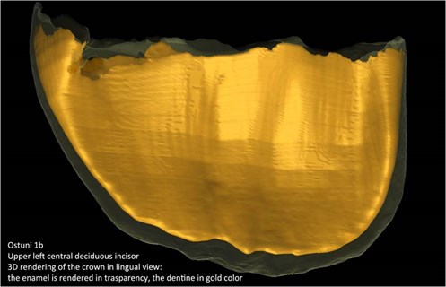
3D rendering of the upper left central deciduous incisor [1] |
 |
|
|
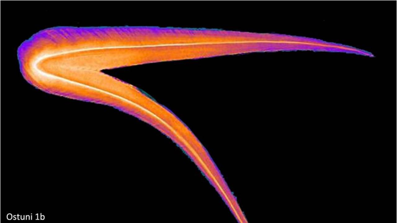
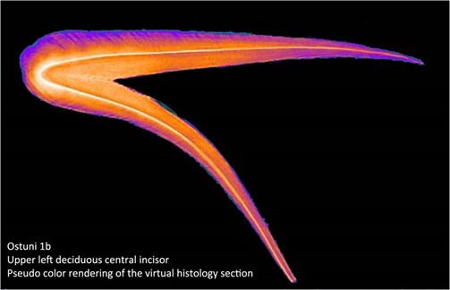
Color rendering of the virtual histology section of an incisor [1] |
 |
|
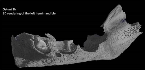
| 3D rendering of the left hemimandible of the fetus [1] |
In an article about the study published by Italian newspaper La Repubblica, Nava declared “The woman’s teeth could provide us information about her diet and her movements but not on her health condition, as they were not growing. Conversely, the milk teeth of the fetus tracks periods of good and bad health while forming, from the 3rd to 4th month of gestation, till they drop. Layers add up daily, which allow us to precisely follow the development of the baby. It’s similar to what happens with tree rings.” [2]
The research team visualized and analyzed three still-forming incisors belonging to the fetus by means of X-ray microtomography. By studying the 108 enamel layers in these milk teeth, scientists could investigate the condition of both the mother and the fetus. Results showed that three severe physiological stresses affecting both individuals occurred during the last two and a half months of pregnancy. This was highlighted by the presence of microscopic stress markers in the dental enamel, which are usually formed after stressful events as a consequence of an altered secretion. These stress episodes could be the symptom of a serious infection that may have led to the death of both the mother and the baby.
The virtual histological analysis has revealed that their death occurred between the 31st and 33rd gestational week and not between the 34th and the 36th, as previous studies had concluded by comparing the skeleton size to today’s fetal growth charts. Being able to precisely establish the gestational age of the fetus allowed researchers to identify some specificities of its embryonal development: “We think in the past a fetus grew faster than today. It may seem counterintuitive, but having a more developed skeleton at the time of delivery could represent an advantage for the baby,” said the researchers from Università Sapienza.
The extraordinary findings were made possible by a fruitful and long-term collaboration between research teams from the Museum of Civilizations and the Dipartimento di Biologia Ambientale of Università La Sapienza in Rome, together with ICTP’s Multidisciplinary Laboratory and the SYRMEP group at Elettra–Sincrotrone Trieste. “Cutting-edge imaging technologies, advanced techniques of data analysis, big data management, and methods for dating samples are all tools physics can offer to archeology,” Tuniz says.
 |
|
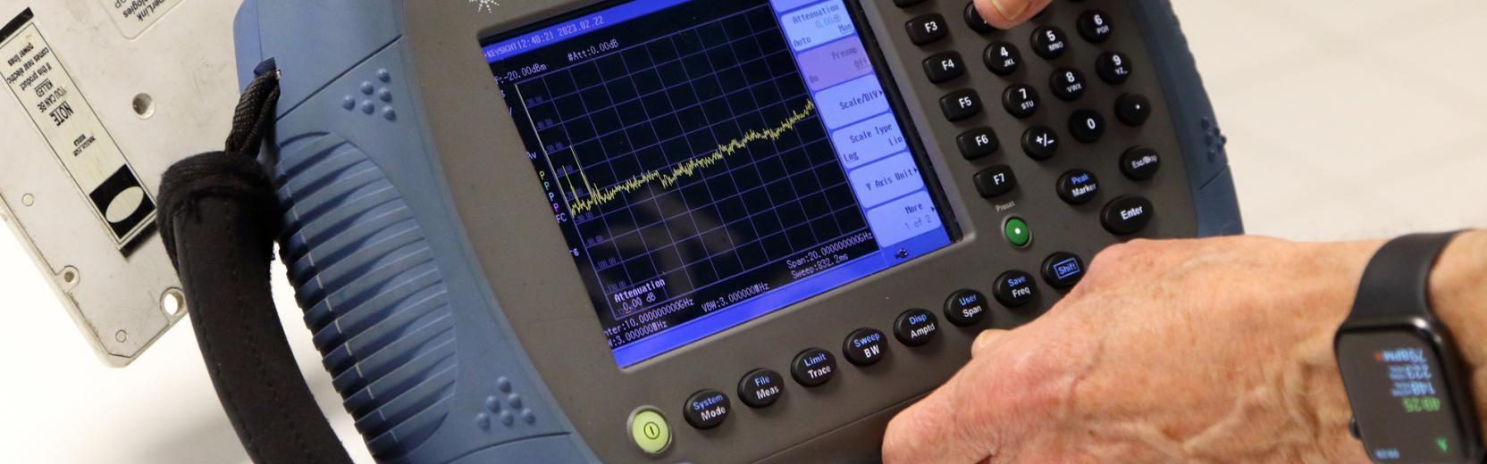
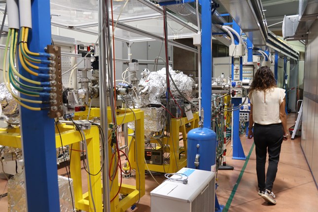
Experimental equipment at Elettra–Sincrotrone, Trieste. Credits: Anna Lombardi |
Scientists at Elettra analyze different kinds of samples, from insects trapped in amber millions of years ago, to ancient musical instruments [3] and fossil bones [4-5]. “To deal with this variety of different specimens, several instruments and methods have been developed at Elettra and ICTP with the aim to exploit all the possible benefits from each one and to cover a large range of possibilities in terms of contrast and spatial resolution, sample sizes and materials to be investigated by X-ray microtomography techniques,” Elettra researcher Lucia Mancini says.
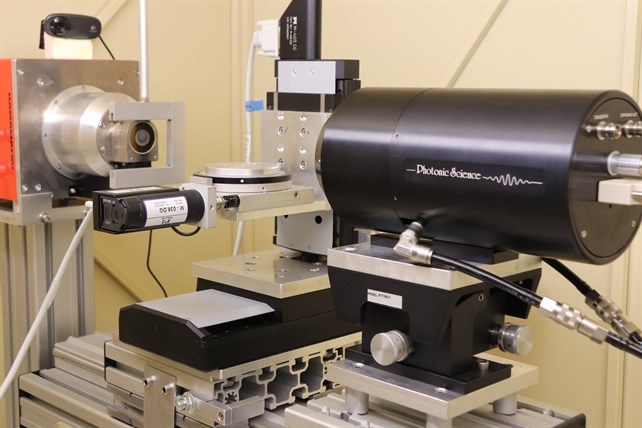 |
| Part of the experimental equipment used to study the fossil teeth. Credits: Anna Lombardi |
The preliminary microtomography studies on the mandible of the fetus realized at the Tomolab's Elettra laboratory, based on a microfocus X-ray source, allowed researchers to study the still-forming incisor contained within it. While the preliminary microtomography studies on the mandible of the fetus allowed researchers to study the still-forming incisors contained within it, the spatial resolution of the 3D reconstruction was not high enough to reveal the finest structures of the dental enamel such as daily and weekly lines.
To achieve that, researchers had to use the unique properties of synchrotron-radiation, available at the SYRMEP beamline of Elettra devoted to hard X-ray imaging, and, above all, to develop a specific methodology of analysis to extract the data and information they needed. The final virtual histological analysis of these precious fossil teeth has revealed, in a non-destructive way, those fine structures of the dental enamel that have been so crucial to get to the results of the study.
“The best part of such a multidisciplinary collaboration,” Lucia Mancini concluded, “is that each researcher contributes the state-of-the-art knowledge of his or her own field and this makes you progress very quickly. It’s a more fascinating and faster process than only reading a book and starting to investigate a field by scratch.”
---- Anna Lombardi
References:
[1] A. Nava et al., Virtual histological assessment of the prenatal life history and age at death of the Upper Paleolithic fetus from Ostuni (Italy), Scientific Reports (2017) doi:10.1038/s41598-017-09773-2
[2] http://ricerca.repubblica.it/repubblica/archivio/repubblica/2017/08/26/…
[3] L. Rigon et al., Synchrotron-radiation microtomography for the non-destructive structural evaluation of bowed stringed instruments, e-Preservation Science (2010)
[4] F. Bernardini et al., Beeswax as Dental Filling on a Neolithic Human Tooth, PLoS ONE (2012) doi:10.1371/journal.pone.0044904
[5] F. Baruffaldi et al., An innovative CCD-based high resolution CT system for analysis of trabecular bone tissue, IEEE Transactions on Nuclear Science (2006)