
seminars
14
Apr
2026
Mathematics
IGAP Special Lecture: Finite multiple zeta values and the poor man's a...
Physical
Back
What information can researchers glean from the 8- to 12-million-year-old fossil remains of apes? Quite a lot, according to Jay Kelley, Adjunct Professor from the Institute of Human Origins at Arizona State University. Kelley has been working with ICTP researcher Federico Bernardini at the centre's Multidisciplinary Laboratory (MLab) to establish if the fossil remains, which were collected from Pakistan and India between 1980 and 2000, relate to the present day orangutan as suggested by some aspects of skull anatomy.
Understanding this relation will help researchers identify patterns of evolution among apes and also understand instances of parallel or convergent evolution in unrelated animals.
Key to their investigation is the use of modern physics tools to uncover the past. The MLab's X-ray microtomography scanner, the only one of its kind in Italy dedicated to paleontology and anthropology, can analyse fossils without damaging them. Among other projects, the scanner has been used in the analysis of a human molar from about 1 million years ago and a piece of humanoid skull that might be the first evidence of Neanderthals in Italy.
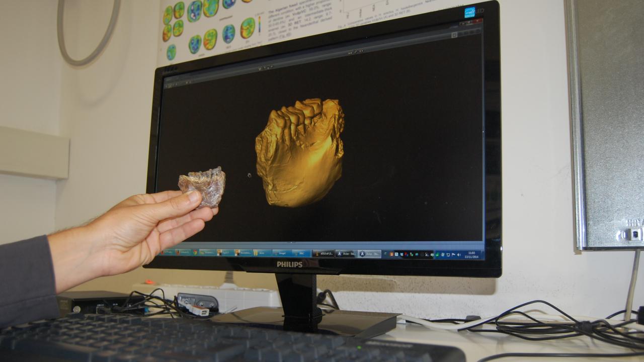
"These [fossil] remains are mostly jaws or teeth, and the teeth preserve a lot of useful information other than just their surface anatomy," says Kelley. "By using an X-ray microtomography scanner, we are able to see the anatomy of the dentin (inner structure of the teeth) that lies below the enamel. While the enamel surface of the molars does not look much like that of living orangutans, the underlying dentine may be more similar to that of orangutans," he explains.
Previous work on the fossil remains has looked at isotope compositions, which become permanently fixed when the tooth forms. Kelley says that sampling the fossil teeth for the composition of the carbon and oxygen isotopes lets researchers understand what the environment would have been like 8 million years ago and what kind of diet the animals followed. For the current analysis at the MLab, the researchers are focussing on understanding how the fossil apes are related to other apes (past and present).
Completing this analysis requires a meticulous process using the MLab's X-ray microtomography scanner. “The scanner is made of three main components—the X-ray tube, sample holder, and a detector," says Bernardini. "To get a virtual rendering of the teeth with all the inner information, we have to first take around 1500 to 3000 radiographs of a single sample by slowly turning it. Then, using specialised software, we produce [virtual] transversal slices of the teeth and all the slices are put together to get an image of the teeth along with their different components like the enamel from the dentin," he explains.
Challenges arise when the dentin and enamel are not sufficiently different in their density, which can happen in cases of fossils. "When fossils lie in the ground for millions of years, minerals might seep into them. If minerals penetrate both the enamel and the dentin to the same extent or make the dentin look as dense as the enamel then you can’t distinguish between the two and you can’t visualise the dentin surface," says Kelley.
In case they do not get a clear picture, there are other possibilities such as analysing some of the teeth at the Synchrotron facilities at Elettra in Trieste. But, Bernardini says that the results using the MLab's X-ray microtomography scanner have been quite good.
Other projects carried out at the MLab include High-Tech Teeth and Northern Italy's First Neanderthal.